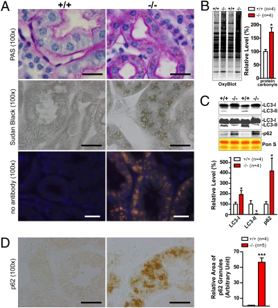
Fig. 3.
Impairment of the autophagy–lysosomal pathway and increases in oxidative damage. (A) Periodic Acid-Schiff staining of cross sections of kidneys shows thicker and darker pink staining in renal tubules and the presence of widely distributed brown granules in boxy cells of renal tubules of 20-month-old LRRK2−/− mice compared with WT controls (+/+). Nuclei were counterstained with hematoxylin (blue). Sudan Black B staining reveals, in renal tubules of LRRK2−/− mice, widely distributed dark brown granules that show bright autofluorescence (orange for merged fluorescence) in the absence of any primary and secondary antibodies. (B) Elevated levels of protein carbonyls (OxyBlot) in Triton X-100–insoluble fractions of kidneys from 20-month-old LRRK2−/− mice. Equal loading of total proteins was confirmed by Ponceau S–staining of the membranes. (C) Western blotting shows dramatically decreased levels of autophagosome marker LC3-II (some LC3-II signals are detected when more proteins were loaded and longer exposure was used) and increased levels of an autophagy substrate p62 in Triton X-100–insoluble fractions of kidneys from LRRK2−/− mice. (D) Immunohistochemical analysis revealed the presence of granular aggregates or inclusions immunoreactive to p62-specific antibody in the deeper layer of the renal cortex of LRRK2−/− mice. Relative areas of granules immunoreactive to p62 antibody were estimated using the ImageJ program (National Institutes of Health). (All scale bars, 20 μm.) Data in all panels are expressed as mean ± SEM. *P < 0.05; ***P < 0.001.

