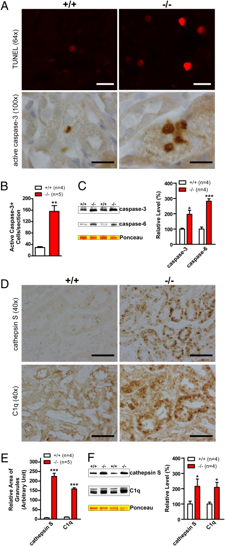
Fig. 4.
Increases in apoptotic cell death and inflammatory responses. (A) TUNEL and immunohistochemical analysis shows increased numbers of TUNEL-positive cells and densely stained active caspase-3–immunoreactive cells, respectively, in kidneys of 20-month-old LRRK2−/− mice. (B) Quantification of active caspase-3–immunoreactive cells shows dramatic increases of apoptotic cells in LRRK2−/− mice. (C) Western analysis shows increased levels of active caspase-3 and caspase-6 in Triton X-100–soluble fractions of LRRK2−/− mice. (D) Immunohistochemical analysis reveals up-regulation of cathepsin S and complement C1q, two commonly used inflammation markers, in LRRK2−/− mice. (E) Relative areas of granules immunoreactive to cathepsin S- or C1q-specific antibody were estimated using the ImageJ program (National Institutes of Health). (F) Western blotting confirms the elevated level of cathepsin S and C1q in Triton X-100–insoluble fractions of kidneys from 20-month-old LRRK2−/− mice. (Scale bars, 20 μm for ×64 and ×100, and 50 μm for ×40.) Data in all panels are expressed as mean ± SEM. *P < 0.05; **P < 0.01; ***P < 0.001.

