Fig. 3.
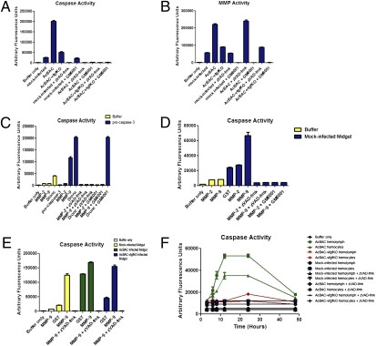
MMPs work upstream of caspase activation and caspases are released into the hemolymph. Larvae were orally infected with AcBAC or AcBAC–vfgfKO, or mock-infected and fed inhibitors zVAD-fmk, GM6001, or DMSO. Insects were dissected at 12 h p.i. or as indicated, and lysates were analyzed for enzymatic activity. (A, B, and E) Midgut lysates were analyzed for caspase (A and E) or MMP (B) activity using DEVD-afc caspase or MMP fluorescent substrates, inhibitors, and proteins. (C) Active MMP or initiator caspase Dronc was incubated with procaspase-3 in the presence or absence of zVAD-fmk or GM6001, and caspase activity measured. (D) Lysates from midguts or buffer only were used to measure caspase activity in the presence of active MMPs. (F) Hemolymph and hemocytes were collected from mock- or virus-infected insects cofed with inhibitors and tested for caspase activity.

