Fig. 4.
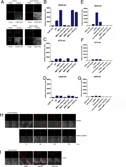
Increased caspase activity is observed during infection with AcBAC. Infected larvae (A) were dissected at 12 h p.i., and TUNEL-positive cells (green) and active caspase (red) were detected. Lysates from uninfected (B–D), virus-, or mock-infected (E–G) midguts were incubated with the effector (DEVD-afc) or initiator (IETD-afc or LEHD-afc) caspase fluorescent substrates and the corresponding caspase inhibitors, DEVD-fmk, IETD-fmk, and LEHD-fmk, and purified proteins. Larvae were infected in the presence or absence of inhibitors or carrier and dissected at the times shown (H) or 12 h p.i. (I), and receptor activation and effector caspase activity were detected. (Upper Left) Red, anti-phosphotyrosine staining; (Upper Right) green, anti-active DrICE staining; (Lower Left) bright field; (Lower Right) merge.

