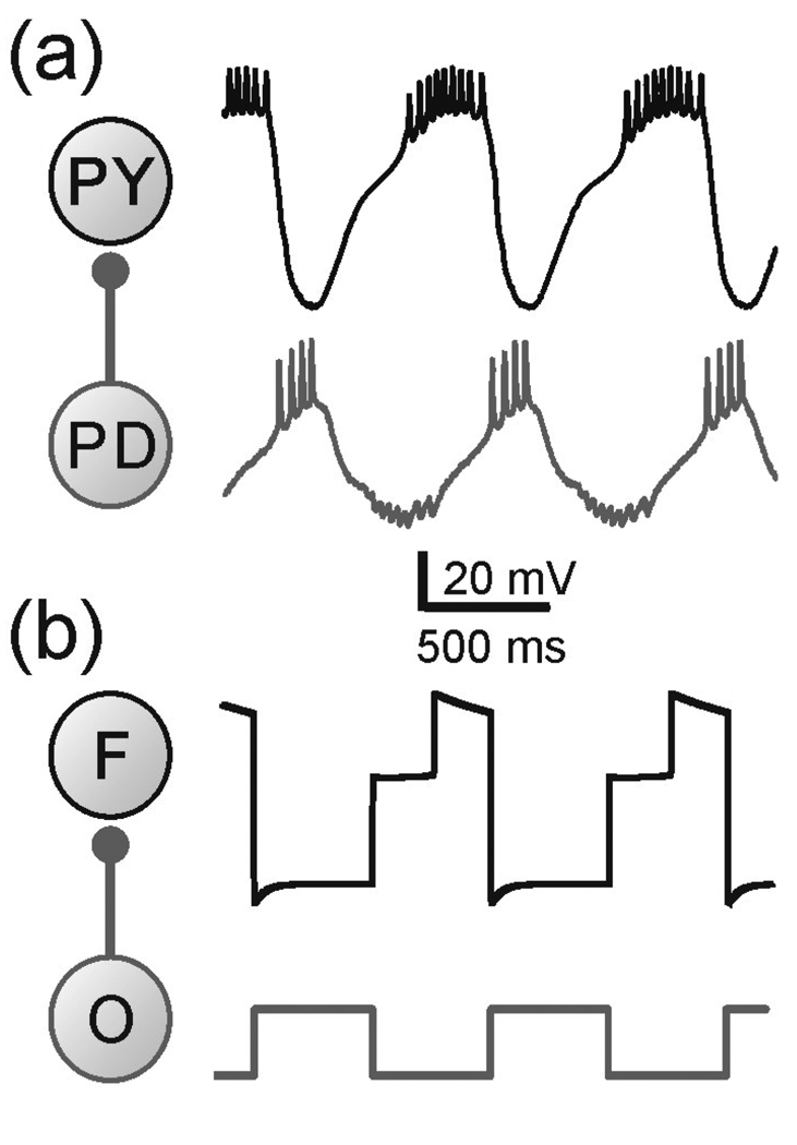
Figure 1.
(a) The network and membrane potentials of the PY and PD neurons in pyloric CPG of the crab Cancer borealis. The follower neuron PY receives inhibitory synaptic input from the pacemaker neuron PD. (b) The network and membrane potentials of the three-variable model. F represents the follower neuron while O the pacemaker neuron or the oscillator. Note that this model represents only the envelope of slow oscillations and the spikes seen in the biological neuron are smoothed over. Model parameters: (in ms) Tin= Tact = 500, τhl = 495, τhm = 810, τhh = 1000, τwl = 40, τwm = 100, τwh = 800; (in mV) EL = −60, ECa = 120, vm = −1.2, km = −18, EK = −84, vw = 15, kw = −5, vn = −6 , kn = −0.5, vh = −10, kh = 0.1, Einh= −80; (in nS) gL = 2, gCa = 4, gK = 8, gA = 4, ginh = 2 ; (in pA) Iext = 75.

