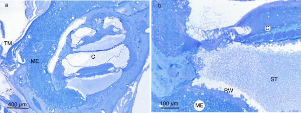
Figure 4.
(A) Middle ear (ME) and inner ear (C) inflammation in dexamethasone-non-responder C3H/HeJ ear. The middle ear inflammatory cells fill the entire space medial to the tympanic membrane. (B) Higher magnification shows inflammatory cells and fibrillar debris in scala tympani (ST). External to the round window membrane (RW) are densely packed inflammatory cells in middle ear (ME). (TM – tympanic membrane)

