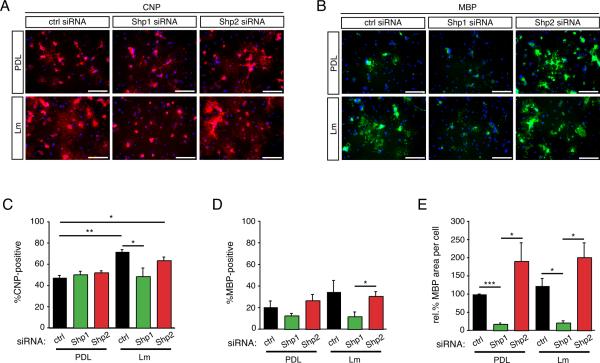
Figure 7. Shp1, but not Shp2, is required for oligodendrocyte differentiation.
Following transfection with Shp1, Shp2, or control (ctrl) siRNA, oligodendrocytes were differentiated for 4 days on either PDL or laminin (Lm) and evaluated by immunocytochemistry to determine the percentage of CNP or MBP positive cells. (A) Representative micrographs of CNP immunocytochemistry (red) and DAPI nuclear stain (blue). Scale bar equals 100 microns. (B) Representative micrographs of MBP immunocytochemistry (red) and DAPI nuclear stain (blue). Scale bar equals 100 microns. (C) Bars represent the mean percentage (± sem) of CNP(+) cells after differentiation on PDL or laminin (Lm). (D) Bars represent the mean percentage (± sem) of MBP(+) cells after differentiation on PDL or laminin (Lm). (E) Bars represent the mean (± sem) relative change in area per MBP(+) myelin membrane sheet after differentiation on PDL or laminin (Lm). Mean areas are normalized to mean area of control cells on PDL (*p<0.05, **p<0.01, ***p<0.001).

