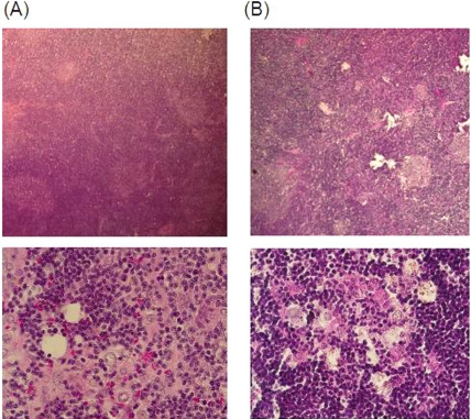
Figure 2.
Microscopic findings following hema-toxylin-eosin staining of BUF/Mna (A) and BUF/ Mna-Rnu/+ (B) thymomas. Under low magnification, enlargement of the cortex area with a reduced medulla is shown. Under high magnification, large-sized round neoplastic thymic epithe-lia associated with abundant lymphocytes can be seen.

