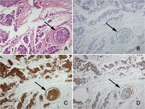
Figure 3.
In situ carcinoma (arrows) surrounded by small cell carcinoma (×200). (A) H&E. Immunohistochemistry for (B) myoepithelial marker p63 confirms the presence of in situ carcinoma, (C) neuroendocrine marker synaptophysin shows positive staining of the in situ carcinoma and the small cell carcinoma, and (D) TTF-1 shows positive staining of the in situ carcinoma and the small cell carcinoma.

