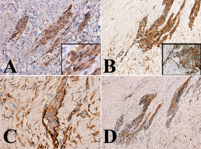
Figure 3.
Representative immunohistochemical staining. Tumor cells were stained with an antibody specific to the vascular endothelial growth factor receptor 3 (VEGFR3) (A), podoplanin (B and C), and CD31 (D). Immunoreactivity with the anti-VEGFR3 antibody was observed in the cytoplasm and nuclei (A, inset). Note the tumor cell clusters in the lymphatic vessels (B and C). The walls of the affected lymphatic vessels were focally obscure; this suggested that the tumor invading to the stroma.

