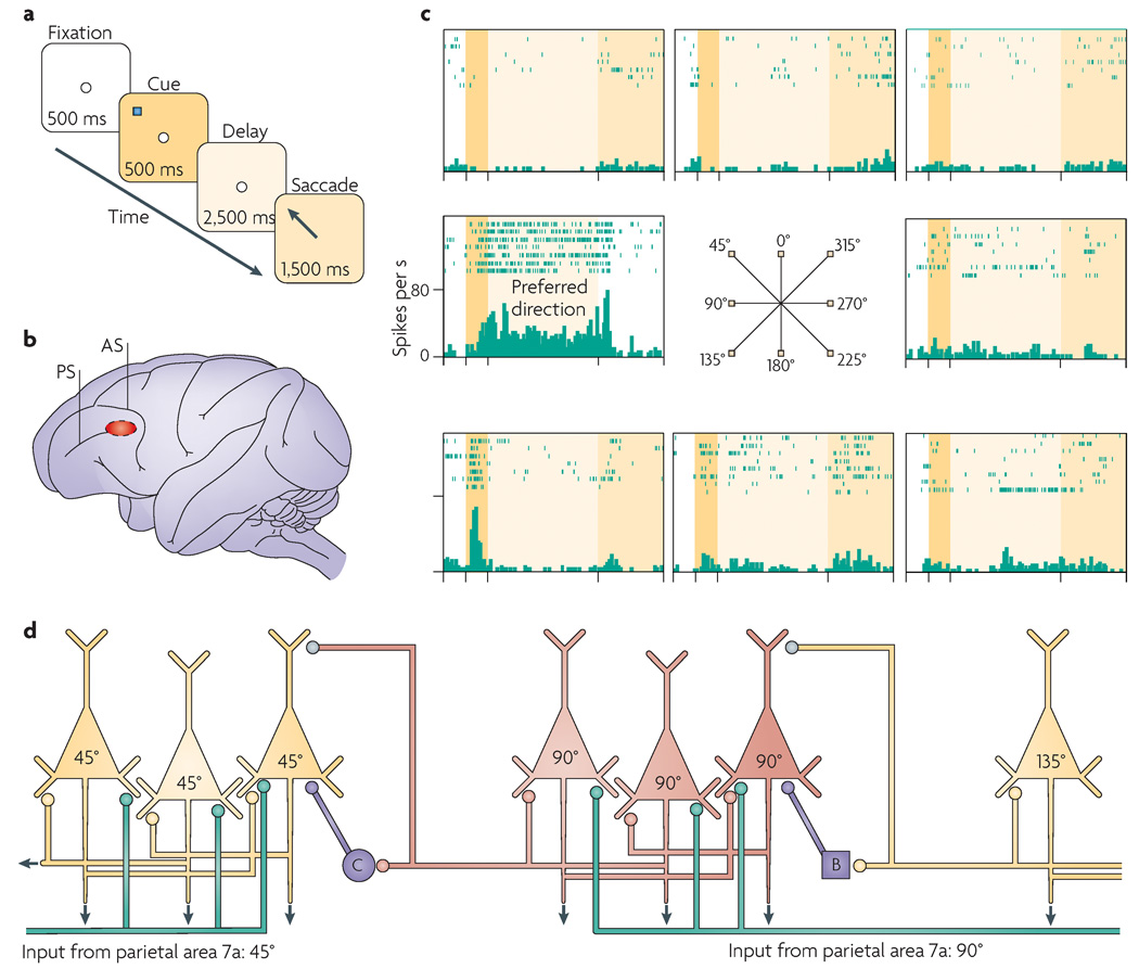
Figure 1. Spatial working memory networks in the dorsolateral prefrontal cortex.
a | The oculomotor delayed response (ODR) task is a spatial working memory task that is used to probe the physiological profiles of prefrontal cortex (PFC) neurons. The subject must remember the spatial position of the most recent cue over a delay period of several seconds and then indicate that position with a saccade to the memorized location. b | The region of the monkey dorsolateral PFC (Walker’s area 46) where neurons show persistent, spatially tuned firing during the delay period in the ODR task. c | An example of a PFC neuron that showed persistent firing during the delay period if the cue had occurred at 90° — the ‘preferred direction’ for this neuron (left middle plot) — but not if the cue appeared in a ‘non-preferred’ direction (other plots). The rasters show the firing of the neuron over seven trials at each spatial position. d | A schematic drawing of the PFC microcircuits that underlie spatial working memory as described by Goldman-Rakic40. Layer III pyramidal cells receive highly processed spatial information (represented by the green lines) from parietal association cortices. Pyramidal cells with similar spatial characteristics engage in recurrent excitation to maintain persistent activity over the delay period (note that the subcellular localization of these excitatory connections is not currently known; they could be on the apical and/or basal dendrites). GABA (γ-aminobutyric acid)-ergic interneurons, such as basket cells (B) and chandelier cells (C) help to spatially tune neurons through lateral inhibition. Inputs from cross-directional microcircuits (neurons with different tuning characteristics) are shown in grey. AS, arcuate sulcus; PS, principal sulcus. Parts a–c are modified, with permission, from REF. 60 ©(2007) Cell Press.

