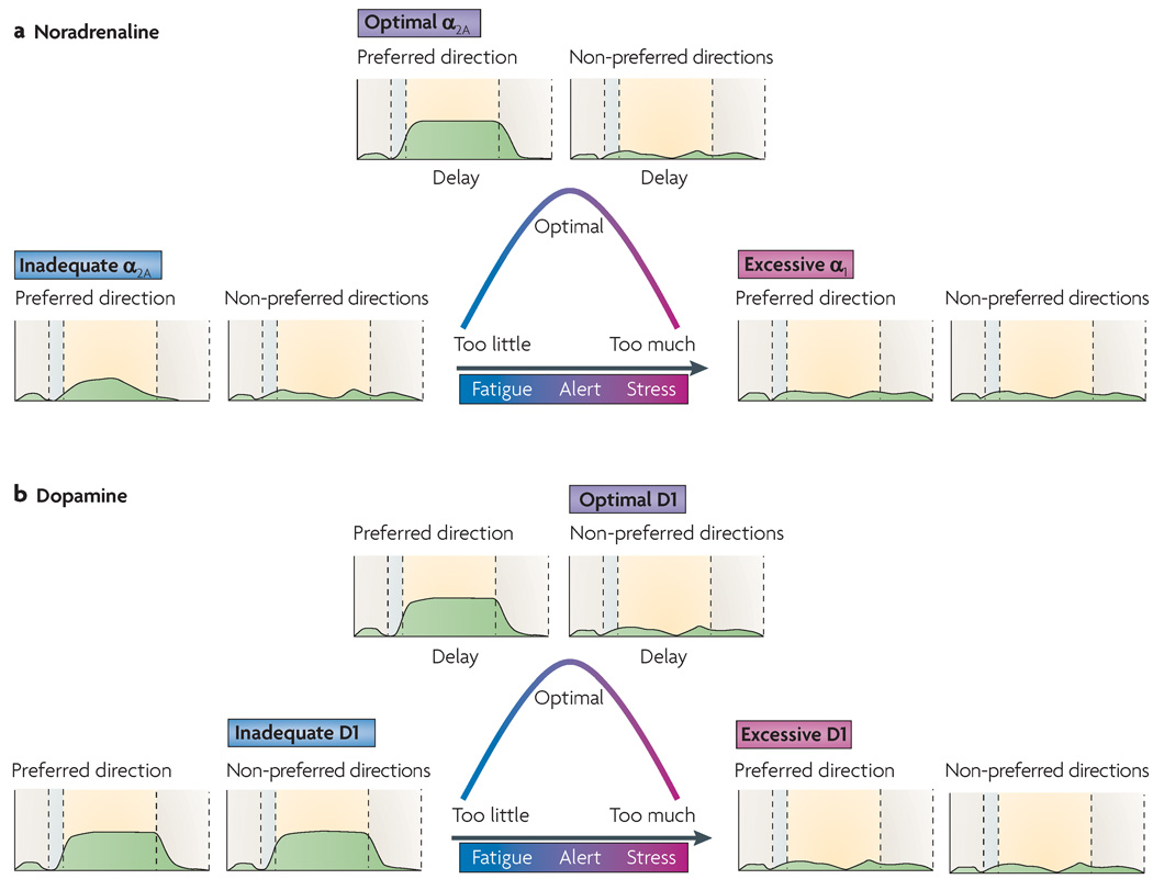
Figure 2. Catecholamine influences on prefrontal cortex physiology and function.
Both noradrenaline (NA, part a) and dopamine (DA, part b) have ‘inverted U-shaped’ influences on prefrontal cortex (PFC) physiology and cognition, whereby either too little or too much of the neurotransmitter impairs PFC function. In an oculomotor delayed response task, with optimal levels of NA or DA release under alert, non-stressed conditions (top of the curves), PFC neurons fire (as shown in the green traces) during the delay period following cues for preferred but not non-preferred directions. NA enhances delay-related firing in response to cues in preferred directions by stimulating α2A-receptors (increasing the ‘signal’), whereas DA weakens delay-related firing in response to cues in non-preferred directions by stimulating D1 receptors (decreasing the ‘noise’). Administration of appropriate concentrations of the α2A-receptor agonist guanfacine or the D1 receptor agonist SKF81297 also has this effect. With high levels of NA release during stress (right side of the curve), NA engages the lower-affinity α1-receptors and so reduces neuronal firing. Similarly, excessive D1 receptor stimulation during stress suppresses cell firing. Administration of the α1-receptor agonist phenylephrine or a high concentration of SKF81297 can mimick the effects of high NA and DA levels, respectively. In each graph, the dotted lines indicate the transition between (from left to right) the fixation period, the cue period, the delay period and the oculomotor response period.

