
Figure 1.
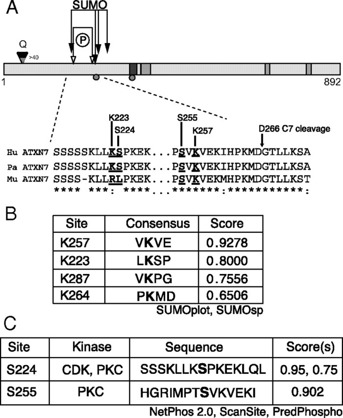
Schematic of the ataxin-7 structure. A, The 892 aa protein contains a nuclear export signal (NES; black rectangle) and three nuclear localization signals (NLS; gray rectangle). The diamond indicates the location of the polyglutamine stretch, and the caspase cleavage sites at Asp-266 and Asp-344 are indicated by circles. Arrows indicate sites of putative PTMs: phosphorylation (open) and SUMOylation (closed). Lysine 257 is indicated with dropped arrow. B, SUMOylation predictions using SUMOplot (Abgent) and SUMOsp (http://sumosp.biocuckoo.org/). Predicted targets shown in bold. C, Phosphorylation predictions using NetPhos2.0 (www.cbs.dtu.dk/services/NetPhos/), ScanSite (http://scansite.mit.edu), and PredPhospho (http://www.nih.go.kr/phosphovariant/html/seq_input_predphospho2.htm). Predicted targets are shown in bold.