
Fig. 3.
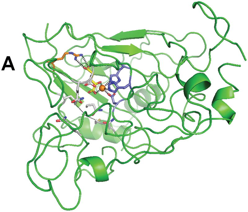
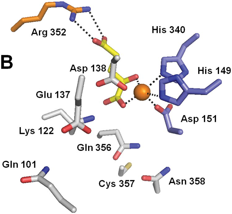
Structure of XanA according to a homology model. (A) The predicted overall fold of the XanA protein is depicted as a green ribbon. (B) A blow-up of the proposed active site. In both panels, FeII is an orange sphere, αKG has yellow carbon atoms, metal ligands have blue carbon atoms, a cofactor-stabilizing residue (Arg 352) is shown with orange carbon atoms, and other postulated active site residues are indicated with white carbon atoms.