
Fig. 7.
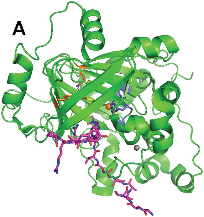
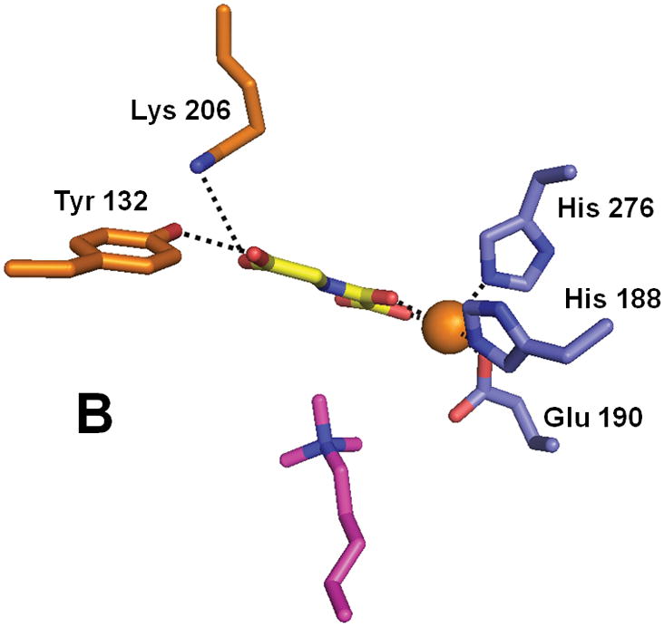
Structure of the histone demethylase JMJD2A (PDB 2p5b). (A) The overall view of the histone demethylase, with bound zinc shown as a grey sphere, the peptide substrate shown with pink carbons, and the αKG homologue N-oxalylglycine replacing the cosubstrate. (B) The active site of JMJD2A with just the trimethyl-Lys portion of the substrate shown. The coloring scheme is the same above.