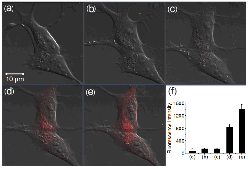
Fig. 4.
Confocal fluorescence images of live human SH-SY5Y cells with the treatment of RS-BE/Fe/ H2O2 (scale bar 10 µm). (a) DIC; (b) the cells incubated with 10 µM RS-BE for 30 min; (c) the cells were then incubated with 10 µM Fe(8-HQ) for 30 min; (d) and (e) the cells were further treated with 100 µM H2O2 for 10 and 25 min, respectively, (f) Integrated emission (547–703 nm) intensity of (a), (b), (c), (d) and (e) images.

