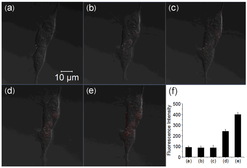
Fig. 5.
Confocal fluorescence images of live human SH-SY5Y cells with the treatment of RS-BE/Cu/ H2O2 (scale bar 10 µm). (a) DIC; (b) cells incubated with 10 µM RS-BE for 30 min; (c) the cells were then incubated with 10 µM Cu(8-HQ) for 30 min; (d) and (e) the cells were further treated with 100 µM H2O2 for 30 and 60 min, respectively; (f) Integrated emission (547–703 nm) intensity of (a), (b), (c), (d) and (e) images.

