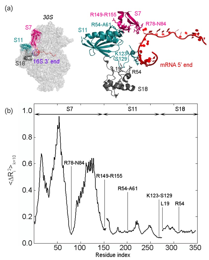
Figure 8.
(a) The 5’ end of the mRNA (red) is surrounded by ribosomal proteins S7 (magenta), S11 (turquoise) and S18 (gray) at the exit channel, shown in two views. The important residues reported in various studies30, 42,43, 84 are shown on the right in stick form. (b) Cumulative mean-square fluctuations summed over the slowest 10 modes 〈ΔRi2〉 k=10 plotted for S7, S11 and S18, and rescaled between 0 and 1 . It is clear that the residues indicated in (a) and (b) surrounding the mRNA undergo extremely small fluctuations in ten slowest most important modes.

