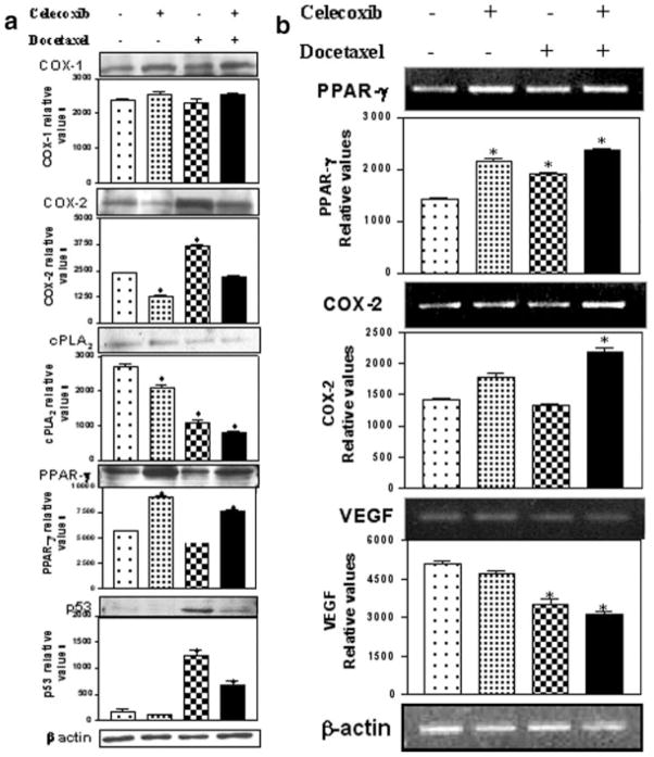
Figure 4.
(a) Western blotting of tumor tissue lysates for COX-1, COX-2, cPLA2, PPAR-γ, p53 and β-actin. The tumors were harvested 28 days post-tumor implantation and lysates were prepared as described in Material and Methods. Protein (30 μg) was loaded in each lane. (b) RT-PCR of COX-2, PPAR-γ and VEGF in tumor tissues. The PCR product sizes for COX-2 (226 bp), PPAR-γ (360 bp), VEGF (121 bp) and β-actin (390 bp) were determined using 100–2680 bp DNA ladder (Fisher Scientific Co., Atlanta, GA). *Significant differences as compared to the vehicle control (p < 0.001).

