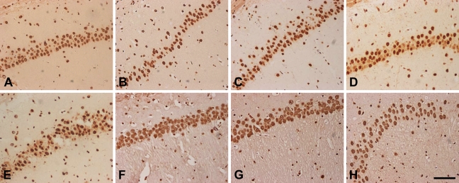
Figure 5.
Immunocytochemistry staining of hnRNP A2/B1 in subfield CA1 of the hippocampus at 3 hr to 72 hr of reperfusion after 120 min of right MCAO, including the sham-operated group (A), and 3 hr (B), 6 hr (C), 12 hr (D), 24 hr (E), 48 hr (F) and 72 hr (G,H) of recirculation. A large number of hnRNP A2/B1-immunoreactive (hnRNP A2/B1-IR) neurons were distributed mainly in the pyramidal cell layer (A). At 3, 6, 12, and 24 hr of recirculation, hnRNP A2/B1-IR expression was reduced after ischemia insults (B–E). At 48 and 72 hr of recirculation, hnRNP A2/B1-IR neurons were increased (F–H). Bar = 100 μm.

