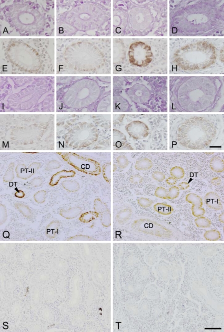
Figure 2.
Light micrographs of the first (A,E,I,M) and second (B,F,J,N) segments of the proximal tubules, distal tubules (C,G,K,O), and collecting ducts (D,H,L,P) in the kidneys of freshwater (A–H)- and seawater (I–P)-acclimated eels. Adjacent sections were stained with periodic acid-Schiff stain (A–D,I–L) and anti-Na+/K+-ATPase (E–H,M–P). Low-magnification views of the kidney sections (Q–T) stained with anti-Na+/K+-ATPase (Q,R) or with the preimmune serum as controls (S,T) in freshwater (Q,S)- and seawater (R,T)-acclimated eels. PT-I, first segment of proximal tubule; PT-II, second segment of proximal tubule; DT, distal tubule; CD, collecting duct. Bars: A–P = 20 μm; Q–T = 50 μm.

