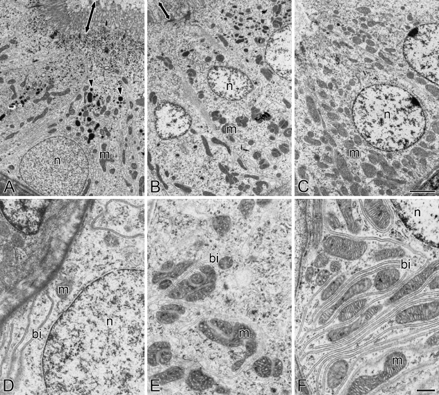
Figure 3.
Transmission electron micrographs of the first (A,D) and second (B,E) segments of the proximal tubule and the distal tubule (C,F) in the kidney of freshwater-acclimated eel. Arrowheads and arrows indicate lysosomal granules and microvilli, respectively. bi, basal infolding; m, mitochondrion; n, nucleus. Bars: A–C = 2 μm; D–F = 500 nm.

