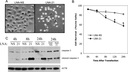
FIG. 4.
Induction of apoptosis upon LNA-21 treatment of cultured murine granulosa cells. Bright field image of rounded up cells after 24 h of treatment with LNA-21 (A). Original magnification ×10. LNA-NS-transfected and LNA-21-transfected cells were double stained with annexin A5 and live/dead violet stain and were analyzed by fluorescence-activated cell sorting 0–24 h after transfection (B). Line graph summarizes the percentage of live cells 0–24 h after transfection, and data (n = 3) were analyzed by t-test; *Means ± SEM are different (P < 0.05) between the LNA-21 and LNA-NS at that time point. C) Representative Western blot (n = 3) of caspase 3 and cleaved (active) caspase 3 in LNA-NS-transfected and LNA-21-transfected granulosa cells 4, 8, and 24 h after transfection, or 24 h after transfection with anti-NS or anti-21.

