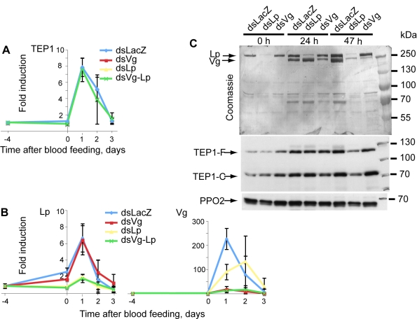
Figure 3. Lp is required for normal Vg expression.
Mosquitoes were injected with dsLp, dsVg, or dsLp+dsVg. (A, B) TEP1, Vg, and Lp expression, respectively, was measured at several time points after P. berghei infection using quantitative RT-PCR. (C) Lp and Vg protein levels in mosquito hemolymph were gauged by Coomassie staining; TEP1 (full length and processed) by immunoblotting. PPO2 served as a loading control. Note that levels of Vg protein are strongly reduced at 47, but not 24, h after infection specifically in dsLp-treated mosquitoes.

