
Figure 4.
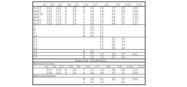
Spacing of conserved cysteines. The protein is indicated on the left (From Table 1) and the cysteine residues are shown on the top. The numbers indicate the number of amino acids between each conserved cysteine. The baculovirus and retrotransposon spacing consensus is shown at the bottom.