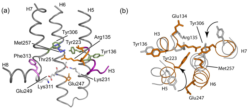
Fig. 16.
Intracellular ionic lock in rhodopsin. Views from the cytosolic surface of the rhodopsin (PDB code 1U19) (a) and opsin (PDB code 3CAP) (b) reveal the disruption of a salt bridge between Arg135 of the conserved E/DRY sequence and a glutamate side chain on H6 at position 247 upon activation. In concert with activation, the side chains of Tyr223 and Tyr306 on helices H5 and H7, respectively, rotate inward into close proximity to the guanidinium side chain of Arg135 as indicated in (b).

