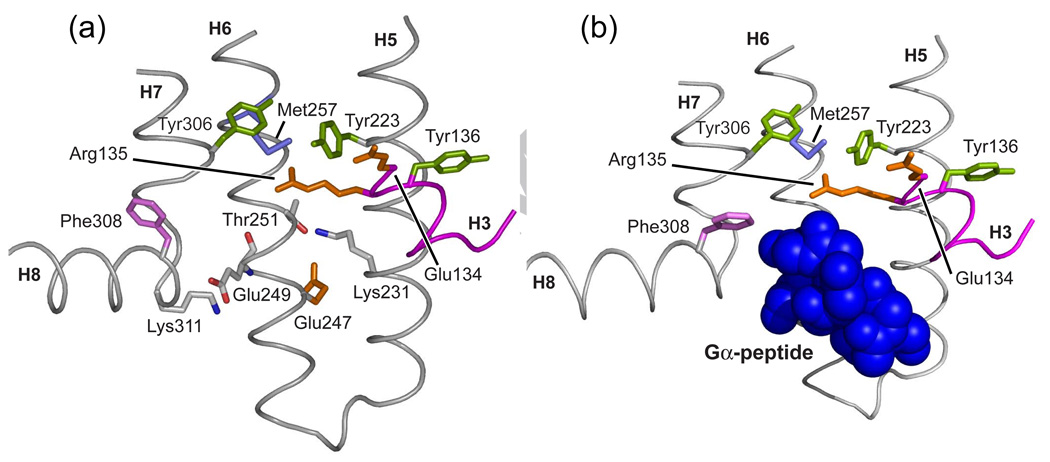
Fig. 17.
G-protein binding site on the intracellular surface of rhodopsin. (a) View of the cytoplasmic region of opsin (PDB code 3CAP) in the region of the intracellular ionic lock. At pH 6.0, the opsin structure exhibits elements of the activated state of rhodopsin. (b) The same view of opsin co-crystallized with an 11-residue peptide corresponding to the C-terminus of transducin (PDB code 3DQB).

