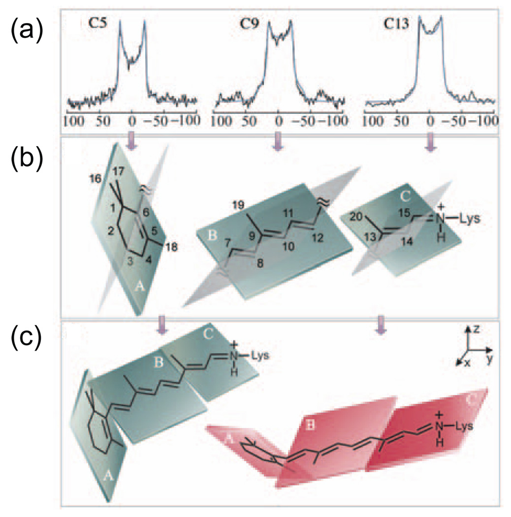
Fig. 6.
Deuterium NMR spectroscopy of the retinal chromophore in rhodopsin. Retinals containing deuterated methyl groups attached to the C5, C9 and C13 carbons were regenerated into rhodopsin. The rhodopsin was reconstituted into POPC bilayers, which were oriented relative to the external magnetic field by ultracentrifugation onto ultrathin glass slides. (a) Experimental and simulated deuterium line shapes are shown for the C18, C19 and C20 methyl groups. The line shapes are sensitive to the orientations of the C5-CD3, C9-CD3 and C13-CD3 bonds relative to the external magnetic field. (b) The C5-CD3, C9-CD3 and C13-CD3 bond orientations can be used to orient specific regions of the retinal chromophore. In these studies, the retinal is divided into three planes containing the β-ionone ring, the polyene chain from C6–C12 and the polyene chain from C13 to the Nε-Lys single bond. Each plane contains one of the deuterated methyl groups. (c) The individual planes are assembled to form a twisted retinal chromophore by allowing rotation about the C5=C6–C7=C8 and C11=C12-C13=C14 torsional angles. The figure is adapted from Ref. [172] with permission from the American Chemical Society.

