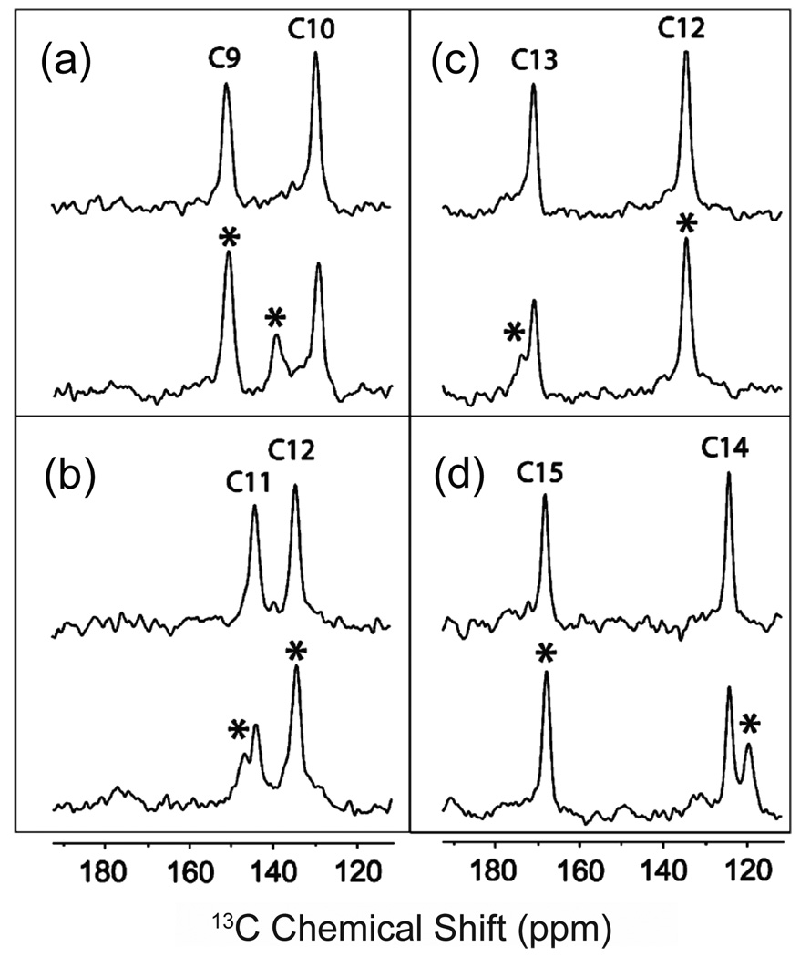
Fig. 7.
Double-quantum filtered 150 MHz 13C NMR spectra of the retinal chromophore in rhodopsin and bathorhodopsin [181]. Double quantum filtering can be used to eliminate the 13C background signal from rhodopsin and the membrane bilayer by inserting directly bonded 13C labels at the C9,C10 (a), C11,C12 (b), C12,C13 (c) and C14,C15 (d) positions of the retinal chromophore. The rhodopsin (top) and bathorhodopsin (bottom) spectra in each panel were both acquired at a temperature <120 K with 7 kHz MAS. The positions of the bathorhodopsin resonances are marked by asterisks. The figure is adapted from Ref. [181] with permission from the American Chemical Society.

