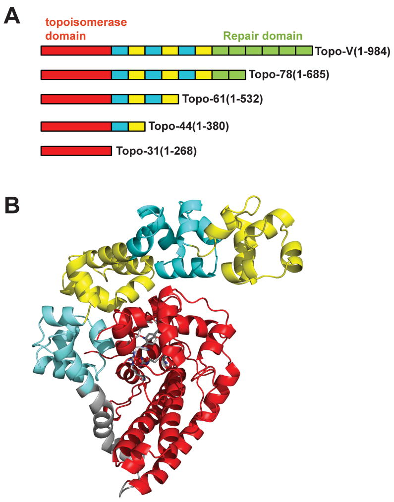
Figure 1. Organization of topoisomerase V.
Topoisomerase V is a multi-domain protein consisting of 24 helix-hairpin-helix (HhH) DNA binding motifs arranged as 12 (HhH)2 domains following the N-terminal topoisomerase domain. A) Schematic diagram of various topoisomerase V fragments. The topoisomerase domain is shown in red, the (HhH)2 domains are shown in alternating colors of cyan and yellow. The (HhH)2 domains with repair activity are shown in green. All fragments shown have topoisomerase activity, but only the full length protein and the Topo78 fragment have repair activity. B) Crystal structure of Topo-61 fragment (Taneja et al., 2006). The coloring scheme is the same as in Figure 1A, except that the linker helix is shown in grey.

