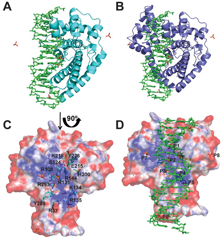
Figure 6. Model showing DNA bound to the topoisomerase domain.
A) Model of a 17-mer double stranded DNA bound to the Topo-31 structure (teal). The DNA is represented as green sticks, where as phosphate ions are represented as orange sticks. DNA binds along the DNA binding groove and five of the eight phosphate ions noted in the Topo-44 structures coincide with the DNA backbone. B) Model of Topo-44 (Form II, B subunit: blue) binding to 17-mer double stranded DNA. Note that the linker helix and the (HhH)2 domains interfere with DNA binding to the topoisomerase domain and are likely to move away to allow binding. C) Electrostatic surface representation of the Topo-31 structure. The positively charged DNA binding groove is clearly visible and the phosphate ions are bound in this groove. The orientation corresponds to a 90° rotation of the one shown in Figure 6A in the direction of the arrow. Note that the DNA binding groove goes from one end of the molecule to the other and it is narrower at one end (start of the linker helix) and wider at the other end. The putative active site residues (green sticks) are located at the wider end of the groove. Other residues lining the groove and interacting with the phosphate ions are shown as cyan sticks. D) Electrostatic surface representation of Topo-31 with phosphate ions (orange) and DNA (green). Three phosphate ions (P3, P4, and P5) coincide with the phosphates of one of the DNA strands, where as P1 coincides with a phosphate of the opposite DNA strand. The model shows that the DNA binding groove of topoisomerase V is wide enough to bind DNA and that the movement of linker helix and (HhH)2 domains are required to accommodate the DNA. The electrostatic potential was calculated with a dielectric constant of 80 for solvent and 2 for protein. The surface is colored with a blue to red gradient from +10 to −10 KbT/ec.

