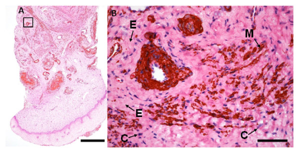
Figure 3.
Immunohistochemical staining of smooth muscle actin in section from the human cervix. (A) Longitudinal section of a biopsy including the epithelium. Bar: 500 μm, (B) a blood vessel surrounded by extracellular matrix (E), smooth muscle cells (M) and connective tissue nuclei (C) (details from boxed-region in A). Bar: 50 μm. Sections were immunostained with an antibody against smooth muscle actin and counterstained with HE.

