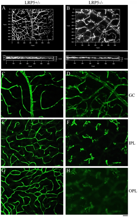
Figure 2. Endothelial cells are unable to form vascular network in the outer- and inner-plexiform layers of LRP5 mutant mice.
A comparison of retinal vasculature visualized by Sca1-GFP positive endothelial cells in 4-week-old LRP5 littermates. 3D images of control LRP5+/− (A) and mutant LRP5−/− (B) are shown. The upper panels are front-views of 3D retinal vasculature and the lower panels are images rotated 90 degrees relative to the upper panels. Single images (C to H) corresponding to the surface vessels (labeled as GC for ganglion cell layer) and vessels in two deeper layers (IPL: inner-plexiform layer; OPL: outer plexiform layer). (C, E, G) images for control LRP5+/− and (D, F, H) for mutant LRP5−/−. Note a complete 3-layer network in the control (A, lower panel) but a lack of vessels in the IPL and OPL of LRP5−/− (B, lower panel). LRP5+/− retina shows normal capillary network in the IPL and OPL (E and G) but LRP5−/− retina displays mainly GFP-positive endothelial cell clusters in the IPL (F) and almost no GFP-positive endothelial cells in the OPL (H).

