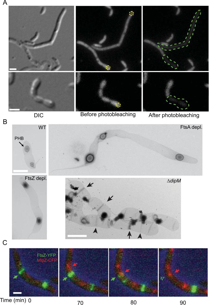
Fig. 2.
Characterization of the cell filamentation phenotype associated with the ΔdipM mutation. (A) FLIP experiment of diffusible cytosolic GFP expressed in ΔdipM cell filament (strain CJW3449) to assess the extent of cell compartmentalization. Yellow dotted circles indicate the regions targeted for photobleaching. The green dotted lines show the extension of the cytoplasmic space as determined by the extent of bleaching.(B) Transmission electron micrographs of isolated sacculi from wild-type (CB15N), ΔdipM (CJW3137), FtsZ-depleted (YB1585) and FtsA-depleted (CJW3187) cells. Depletion was achieved by growing cells in the absence of xylose (inducer) for 5 h (FtsZ) or 8 h (FtsA). In the wild-type panel, the arrow points to a PHB (polyhydroxybutyrate) granule trapped in the sacculus. In the ΔdipM panel, the arrowheads and arrows indicate PG-rings and SP-rings, respectively. (C) Selected frames from a time-lapse microscopy sequence of a ΔdipM cell filament (CJW3448) expressing FtsZ-YFP (green) and MipZ-CFP (red). ftsZ-yfp expression was induced by the addition of 0.3% xylose for 3 h prior to the start of the time-lapse experiment. In all panels, the scale bars correspond to 1 μm.

