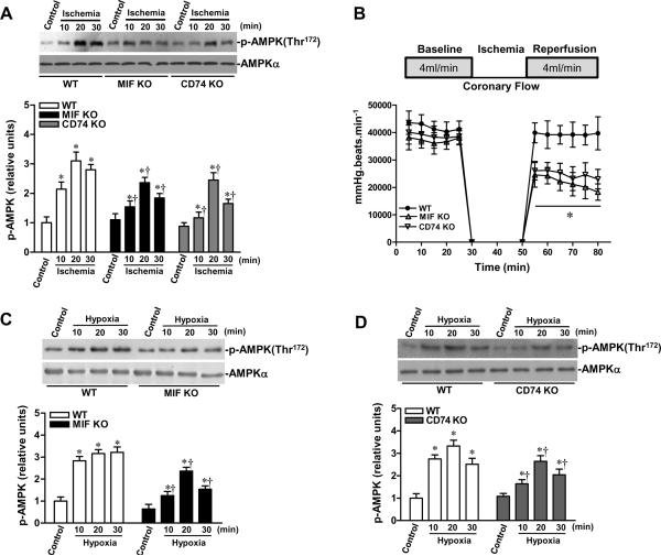
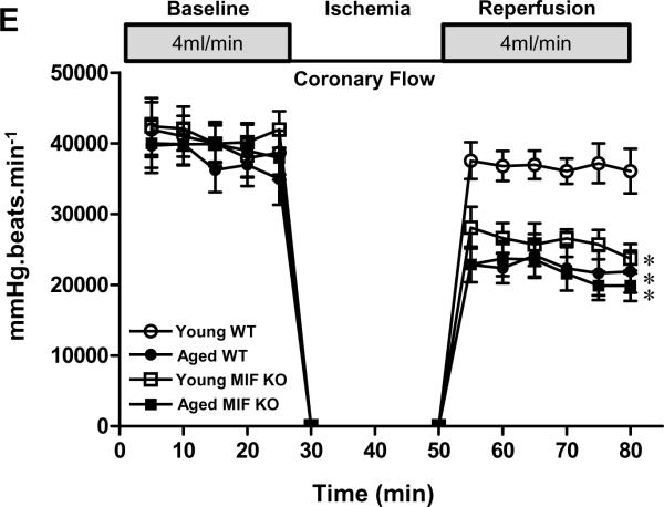
Figure 5.
Impaired AMPK signaling in MIF KO and MIF receptor (CD74) KO hearts. A, MIF KO, CD74 KO and WT mice were subjected to in vivo regional ischemia by LAD occlusion for either10, 20, or 30 min to determine the degree of ischemic AMPK activation (upper panel). Bars represent the relative levels of p-AMPK (lower panel). n=6 per group, *P<0.01 vs. control, respectively; †P<0.05 vs. WT ischemia, respectively. B, Heart rate-left ventricular pressure product of isolated WT, MIF KO and CD74 KO hearts, n=4 per group, *P<0.05 (both MIF KO and CD74 KO) vs. WT. C and D, The kinetics of AMPK phosphorylation induced by hypoxia in WT, MIF KO and CD74 KO cardiomyocytes, n=6 per group, *P<0.05 vs. control, respectively; †P<0.05 vs. WT hypoxia, respectively. E, Heart rate-left ventricular pressure product of isolated young and aged WT hearts, young and aged MIF KO hearts, n=4 per group. *P<0.05 vs. young WT, respectively.


