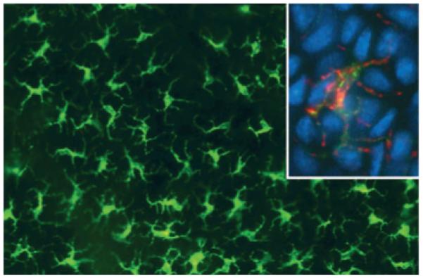
Fig. 1.

Langerhans cells in an epidermal sheet from mouse skin. They are visualized by immunostaining with anti-major histocompatibility complex (MHC)-II antibodies in the large picture. The inset depicts double-labeling of MHC-II (green fluroescence) and langerin (red fluorescence). Nuclei are counterstained with DAPI (4′,6-diamidino-2-phenylindole) (blue). Conventional epi-fluorescence microscopy.
