
Fig. 2.
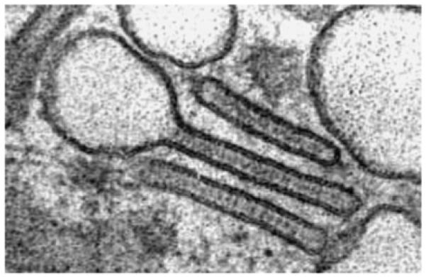
Transmission electronmicroscopy of Birbeck granules in Langerhans cells from human epidermis. Note the conspicuous rod and tennis racket shapes, and the very precisely ‘drawn’ limiting membranes of the structures. The diameter of the rod portion is about 50 nm.