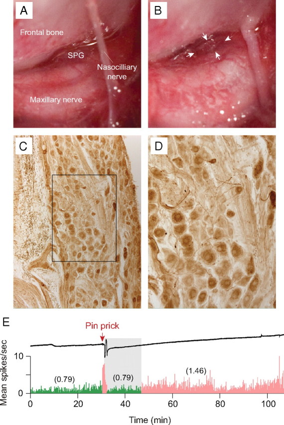
Figure 7.

Effects of ipsilateral SPG ablation on sustained neuronal activation. A, Surgical preparation exposing the rat SPG and adjacent structures. B, The same preparation after removal of the SPG. C, D, Immunostaining for vasoactive intestinal peptide in the isolated SPG (D shows higher-power detail of the boxed area in C). E, An example of a CSD wave induced by mechanical stimulation of the cortex (upper trace) that was associated an initial burst of neuronal firing, followed by delayed onset (gray area) of sustained activation (bottom trace). Note that neuronal activity (numbers in parentheses) after CSD (pink) was approximately twofold higher than baseline activity (green). Bin width = 10 s.
