
Figure 1.
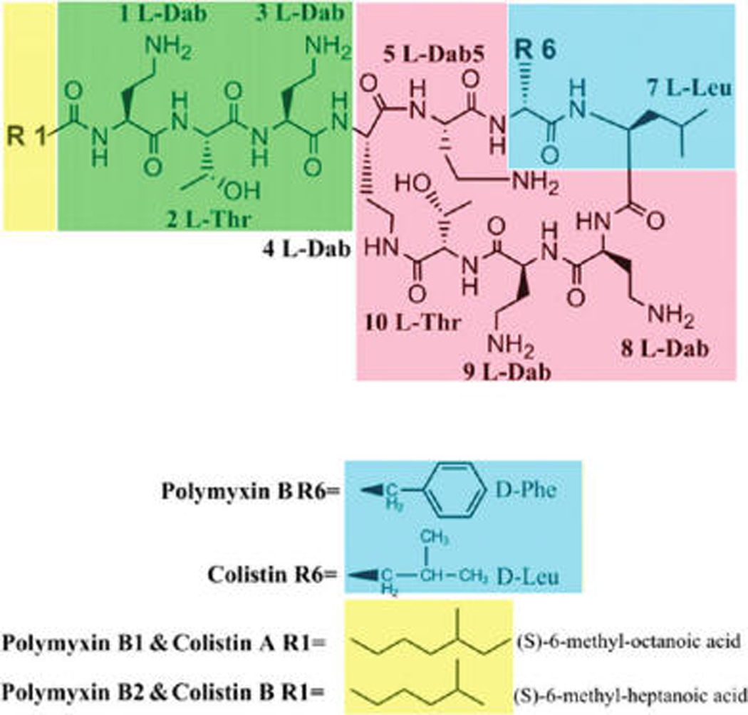
Chemical structures of polymyxin B and colistin. The functional segments of polymyxins are colored as follows: yellow, Na fatty acyl chain; green, linear tripeptide segment; red, the polar residues of the heptapeptide; blue, the hydrophobic motif within the heptapeptide ring. The amino acids positions are numbered in accordance to references in the text.