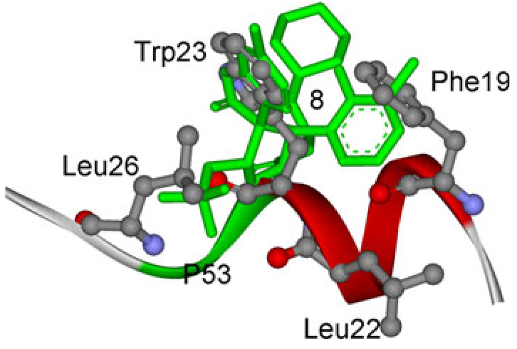
Fig. 12.
Superposition of the averaged structure of compound 8 to the p53 peptide conformation in the crystal structure of p53 peptide conformation in complex with MDM2. Four residues, Phe19, Leu22, Try23, and Leu26 in ball and stick representation. The compound 8 is colored in green with stick representation

