Figure 2.
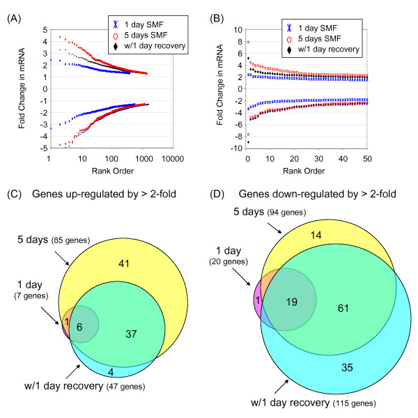
mRNA profiling of SMF-treated hEBD cells. (A) The magnitude and number of genes that showed statistically significant changes in mRNA levels after SMF exposure compared to untreated control cells are given (four data points, representing genes with ≥ 5-fold changes in mRNA levels are not indicated in (A) but are shown in the expanded view of the 50 highest up- and down-regulated genes provided in (B); the "missing" genes are listed in Tables 2–4). Venn diagrams depicting the number of genes up- or down-regulated by ≥ 2-fold compared to cells continuously incubated under normal culture conditions are shown in Panels C and D, respectively. Note that the "1 day SMF" designation refers to the Group 2 treatment conditions described in the Methods section, (the greatest up- and down-regulated genes are listed in Table 2); "5 days SMF" refers to Group 3 cells (Table 3); and "w/1 day recovery" refers to Group 4 cells (Table 4); in all cases comparison is made to Group 1 control cells that were not exposed to SMF.

