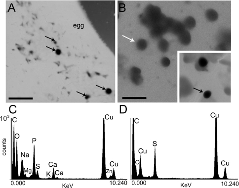
Figure 2. Electron microscopy and X-ray microanalysis of whole intact eggs and homogenates.
(A and B) Electron energy-filtered transmission electron microscopy of an unfixed and unstained egg (A) or an egg homogenate (B) from A. punctulata. Dense granules are identified by black arrows. (C) Typical X-ray microanalysis spectrum of dense granules found in egg homogenates [black arrow in (B)]. (D) Typical X-ray microanalysis spectrum of a less-electron-dense vesicle (probably a yolk platelet) [white arrow in (B)] found in egg homogenates. Scale bars in (A) and (B) are 3 μm.

