Fig. 3.
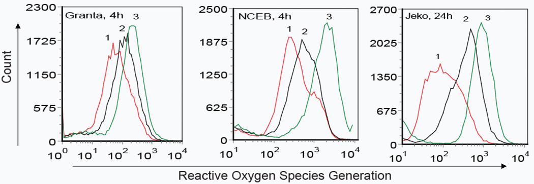
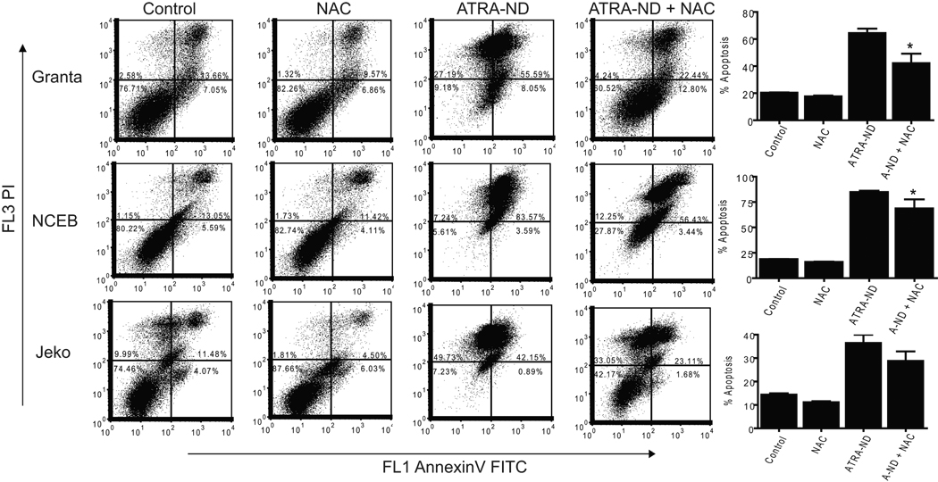
ATRA-mediated ROS generation in MCL cells. (A) Granta, NCEB, and Jeko cells were treated with medium alone (Peak 1), 20 µM naked ATRA (Peak 2) or 20 µM ATRA-ND (Peak 3) for 4 h (Granta, NCEB) or 24 h (Jeko) at 37 °C. ROS production in live cells was measured by flow cytometry as described in Materials and Methods In all 3 cell lines, there is an increase in ROS after ATRA-ND (peak 3) compared with naked ATRA or medium alone. (B) Incubations as in A, except an additional incubation of 20 µM ATRA-ND (A-ND) plus 10 mM N-acetylcysteine (NAC) was included. After 48 h, cells were analyzed for apoptosis as described in Materials and Methods. Early and late apoptotic percentages (AnnexinV/PI positive) from each dot plot were combined to represent total apoptosis (summarized in the bar graphs). In all 3 cell lines, there is an increase in apoptosis after ATRA-ND, which is abrogated by NAC. Values shown are the mean ± SD (n = 3). *p<0.01 Vs. ATRA-ND


