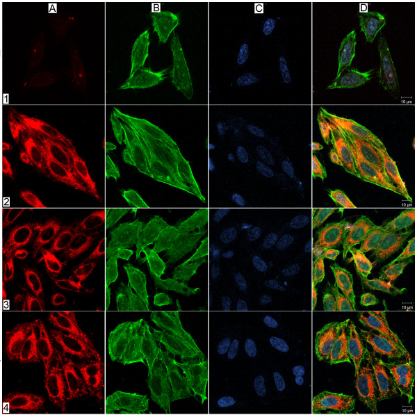
Figure 1.
Infection of U. diversum in HEp-2 cells. LSCM optical sections showing internalization of U. diversum in HEp-2 cells after 1 minute (1), 30 minutes (2), 3 hours (3) and 12 hours (4) post-infection. Ureaplasmas were labeled with Vibrant Dil (in red, A), HEp-2 actin filaments stained with phalloidin-FITC (in green, B) and Hep-2 nuclei stained with TO-PRO-3 (in blue, C). In D, merging images A, B, and C. One minute after infection, ureaplasmas were observed inside HEp-2 cells, and after 30 minutes the presence of ureaplasmas inside cells increased. After 3, 8 and 12 hours of infection, ureaplasmas were observed throughout cells cytoplasm.

