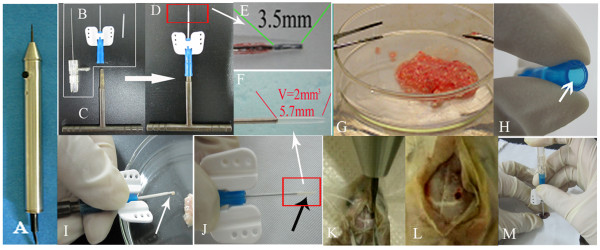
Figure 1.
Illustration of nude mice orthotopic transplantation with glioma tissue. A: micro-skull drill; B: trochar; C tissue propeller; inset in D and E: the depth of injection into mouse brain; G comminuted tumor tissue; H put some tissue into the rear part of trochar (see arrow); I: tumor tissues was packed to the trochar cannula with propeller for transplantation, superfluous tumor tissue were overflowed from the distal end of trochar under the pressure of propeller (see arrow);F and inset in J: exactly 2 mm3 tumor tissue lefted for transplantation (see black arrow); K: drill the hole; L:the burr hole; M: the tumor tissue (J) was injected slowly into brain via the hole (I), then pulled out the trochar slowly, sealed the hole with bone wax and sutured the scalp.

