
Figure 4.
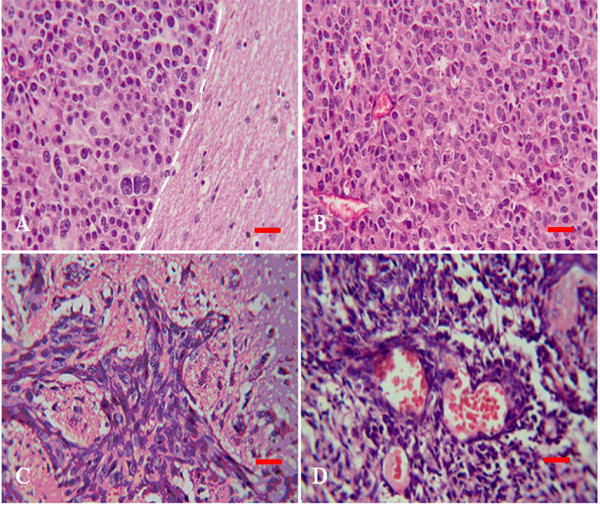
Transplantation tumor observed by HE staining. Tumor cells of brain metastasis (A and B) were small round, easy to see caryocinesia, rare to see multinucleated giant cell and did not form glandular cavity in somewhere (B). Boundary (white dash line) between tumor (left) and normal brain tissues (right) was very clear (A). There was no apparent boundary can be seen between glioma tumor and surrounding brain tissues (C and D) and tumor cells invaded like chicken wire. Tumor cells were fusifirm, star-like, triangle and so on. Abundant vessels shown in tumor tissues and the dndothelial cells were hyperplasy (D).