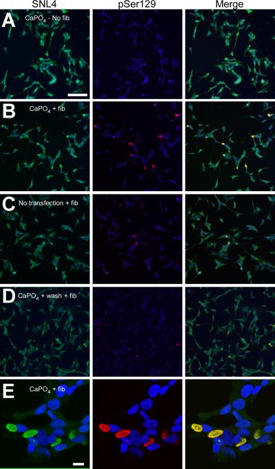
Figure 5.
Double-immunofluorescence of SH-SY5Y neuroblastoma after recombinant α-syn fibril mix treatment. Double-immunofluorescence was performed with SNL4 (green) and pSer129 (red) on SH-SY5Y neuroblastoma that were stably transfected with WT α-syn. Cells were treated with calcium phosphate precipitation (CaPO4) using the control pcDNA3.1 plasmid (A,B,D,E) and/or recombinant 21–140 α-syn fibril mix (fib) (B–E). Removal of CaPO4 (wash), followed by fibril mix treatment was also investigated (D). α-Syn aggregates were formed when cells were treated with recombinant α-syn fibril mix; however, a significant increase in the propensity to form aggregates was observed with concomitant treatment of both CaPO4 and fibrils. (E) Confocal microscopy image of α-syn aggregates in SH-SY5Y neuroblastoma. Bar scale = 100 μm for A–D; 10 μm for E.

