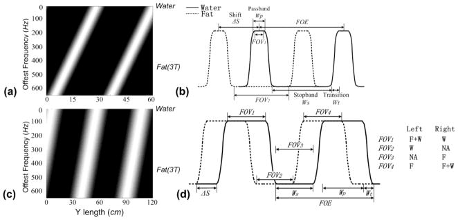
Fig. 2.
(a) The spatial shift of the spin excitation profiles along the phase encoding direction (along the horizontal) as it varies with the amount of off-resonance (along the vertical) for a 2DRF pulse; (b) A 1D idealized plot of the separation of fat and water excitation profiles which enable both reduced FOV imaging and fat suppression as with the π method; (c) The spatial shift of the spin excitation profiles as in (a) for a 2DRF pulse for spatially varying fat-water excitation; (d) A 1D idealized plot of the separation of fat and water excitation with a 2DRF pulse as in (c) that gives the potential for variable fat-water distributions within the FOV as, for example, can be achieved within FOV1-FOV4. Ws: the stopband width, Wt: the transition width, Wp: the passband width, FOE: the field of excitation, and ΔS: the spatial shift.

