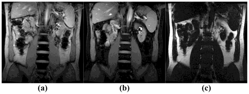
Fig. 4.

Full FOV fat-water separation was performed on a healthy volunteer at 3T (TE=8ms, TR=14ms, flip angle=45°, FOV=34cm, matrix size=256 × 256, slice thickness=8mm). (a) Reference image produced by the original SINC pulse in the product FGRE sequence; (b) A water-only image was obtained by a 5 × 720 μs SPSP pulse; (c) A fat-only image was obtained by the same SPSP pulse;
