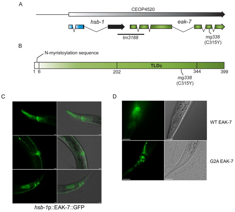
Figure 5. eak-7 gene structure, protein domain organization, and expression pattern.
A. eak-7 gene structure. eak-7 is the downstream gene in an operon with hsb-1. The deletion in the eak-7(tm3188) allele begins 249 bp upstream of the eak-7 initiator methionine codon and deletes portions of exons 1 and 2 of eak-7. The mg338 allele is a C/T transition that results in a cysteine-to-tyrosine mutation at amino acid 315 (C315Y). B. EAK-7 domain organization. The N-myristoylation motif and TLDc domain are shown. C. hsb-1p::EAK-7::GFP expression pattern in L4 larvae. hsb-1p::EAK-7::GFP is strongly expressed in the pharynx and nerve ring (top panels), the vulva and ventral nerve cord (middle panels), and in intestinal cells and cells surrounding the anus (bottom panels). Images were taken at 400× magnification. The scale bar is 10 μm. D. Subcellular localization of wild-type EAK-7::GFP (top panels) and EAK-7::GFP harboring a mutation of glycine 2 to alanine (G2A; bottom panels) at 1000× magnification. The scale bar is 10 μm.

